
Figure 2.
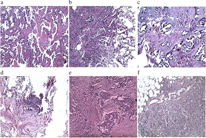
Hematoxylin and eosin (HE) staining of the nodule (a) in the apical segment and (b) the anterior segment of the upper lobe of the right lung. Adenocarcinoma infiltration was observed in the lung tissue, and acinar was the dominant type. (c) HE staining of the subpleural nodule slice in the lower lobe of the right lung. Inflammatory cell infiltration was observed in the lung tissue. (d) HE staining of the paravertebral nodule slice in the lower lobe of the right lung. Adenocarcinoma infiltration was observed in the lung tissue, and acinar was the dominant type. (e) HE staining of the lump slice in the right breast, with the breast infiltrating ductal carcinoma. The lesion was ductal carcinoma in situ. (f) HE staining of the lymph node slice in the right armpit.