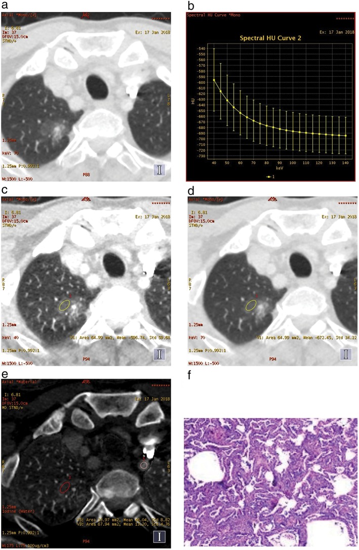
Figure 1.

A mixed ground glass nodule in (a) the right upper lobe, (b) the spectral curve, (c) the monochromatic computed tomography (CT) number obtained at 40 keV and (d) 70 keV energy levels on transverse monochromatic CT image, and (e) iodine concentrations of the lesion and the left subclavian artery on iodine‐based material decomposition images, obtained from single spectral CT acquisition (section thickness 1.25 mm) in the arterial phase. (f) Lepidic predominant adenocarcinoma in a 54‐year‐old man was confirmed by postoperative pathology.
