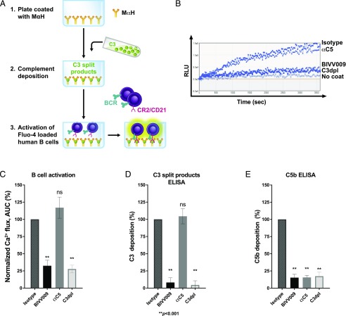
FIGURE 2.
C1s inhibition prevents complement-enhanced BCR activation in primary human B cells. (A) Schematic of an in vitro assay of B cell activation by BCR ligands and deposited complement. (1) Plates are coated with mouse anti-human Ab. (2) Complement is deposited on mouse anti-human–coated plates from either 5% NHS treated with 100 μg/ml human IgG4 (isotype), 100 μg/ml BIVV009 or anti-C5 Ab, or from 5% human C3dpl. (3) Activation of B cells loaded with Fluo-4 stain in mouse anti-human plates with deposited complement is measured as 488 nm fluorescence in a 96-well plate reader. (B) Representative Ca2+ flux traces of Fluo-4–stained primary normal human B cells incubated in mouse anti-human plates treated as described in (A). No coat refers to cells incubated in uncoated wells. (C) Normalized Ca2+ flux in cells from (B) (normalized to isotype control as in Fig. 1E). Data are mean values from independent experiments on B cells from 10 separate donors. (D) C3-split products ELISA of plates from (B). (E) C5b ELISA of plates from (B). Error bars are SEM. Statistics are one-way ANOVA comparison with isotype control. **p < 0.001. αC5, anti-C5; MαH, mouse anti-human.

