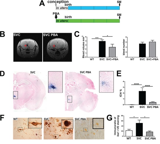
Figure 1.

PBA reduces ICH. (A) Overview of preventative PBA treatment from conception to point of analysis. (B) MRI image of untreated and treated small with vacuolar cataracts (SVC) mice showing ICH (red arrow). (C) Image analysis of MRI data reveals reduced ICH bleed volume (left graph) but not ICH number of bleeds (right graph) (WT n = 6, SVC n = 10, SVC PBA n = 6). (D) Perl’s staining of brains from 5-month-old untreated Col4a1+/SVC and Col4a1+/SVC treated from conception (blue staining). (E) Image analysis of Perl’s staining ICH (WT n = 7, SVC n = 6, SVC PBA n = 12). (F) Immunostaining against Iba1 (brown) on brain sections with detail of dashed square provided in a small square. (G) ImageJ analysis of staining is provided in graph (WT n = 6, SVC n = 5, SVC PBA n = 3). One-way analysis of variance (ANOVA) with post hoc test [Bonferroni (G), Tukey (E); *P-value < 0.05; ***P-value < 0.001].
