Figure 5.

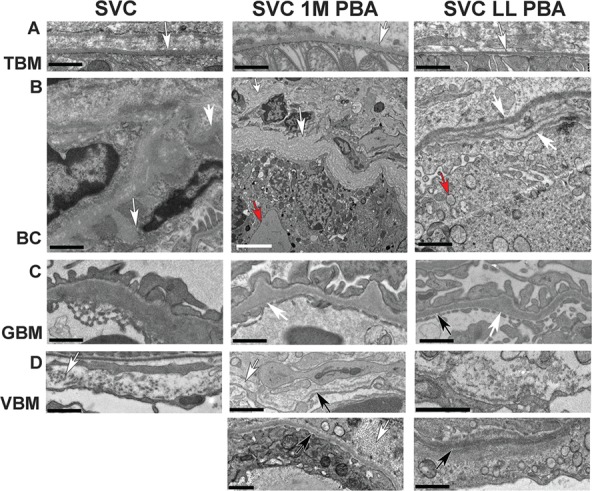
Effect of PBA on BM structure. (A) Normal appearance of BM of tubules (white arrow) in untreated Col4a1+/SVC (SVC) and mice treated for 1 month at 4 months of age (SVC 1M PBA) or from conception for 5 months (SVC LL PBA). (B) Severe defects in BM of Bowman’s capsule in all mice including bulges (white arrow SVC), basket weave appearance (white arrow SVC 1M PBA) and multiple layers (white arrow SVC LL PBA). Evidence of enlarged ER (red arrow SVC 1M PBA) and increased vesicles (red arrow SVC LL PBA) is also observed. (C) Irregular thickening (white arrow) of GBM in treated and untreated mice. Thinner BM areas are also observed (black arrow). (D) VBM defects include interruption (white arrow SVC, 1M PBA), presence of collagen fibrils (white arrow bottom panel 1 M PBA) and more fuzzy but continuous BM (bottom panel LL PBA) black size bar 1 μm, white size bar 5 μm. One-way ANOVA post hoc Tukey test *P < 0.05, **P < 0.01, ***P < 0.001.
