
Figure 2.
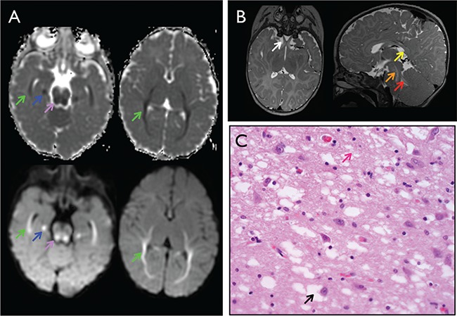
Brain imaging and histology for YARS c.499C > A, p.Pro167Thr homozygotes. (A) Brain MRI at 6 months of age with multiple areas of dysmyelination. Axial diffusion weighted imaging (bottom) and apparent diffusion coefficient (ADC) mapping (top) with restricted diffusion along the white matter of the temporal, occipital and parietal lobes (green arrows), medial temporal lobe (blue arrows) and central tegmental tracts (purple arrows). (B) Abnormal T2 hyperintensity of the optic nerves (white arrow), tegmental tracts (orange arrow) and superior cerebellar peduncles (red arrow) and absent myelination of the corpus callosum (yellow arrow). (C) Hematoxylin and eosin staining of brain biopsy with diffuse vacuolar changes in the gray matter neurons (black arrow) and neurofilament (pink arrow).