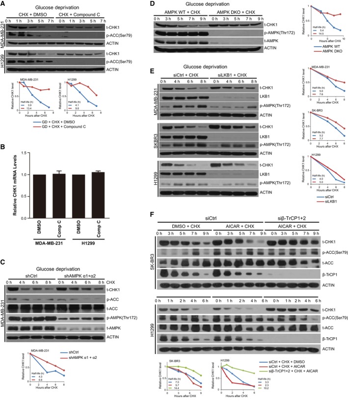
Figure 4.

Inhibition of AMPK blocks glucose deprivation‐induced CHK1 degradation. (A) Indicated cells were grown in glucose‐containing media and treated with or without Compound C at 20 μm (MDA‐MB‐231 cells) or 40 μm (H1299 cells) for 1 h, and then transferred into glucose‐free media with Compound C at the dose indicated above for 3.5 h, upon which time CHX was added to 100 mg·mL−1 for the indicated time points. Cells were harvested and lysed, and protein was separated on SDS/PAGE and immunoblotted for CHK1, p‐ACC, and ACTIN. (B) Cells treated as in (A) were harvested prior to CHX treatment, and RNA was extracted and subjected to RT‐PCR for Chek1. Data generated from three independent replicates; error bars represent SEM, and a paired two‐tailed Student's t‐test was used to assess statistical significance. (C) MDA‐MB‐231 cells were transfected with shCtrl or a mixture of shRNAs targeting AMPK α1 and AMPK α2. Cells were transferred to glucose‐free media and harvested at the indicated time points following addition of 100 mg·mL−1 CHX. Cells were harvested and lysed and protein separated on SDS/PAGE and immunoblotted for CHK1, p‐ACC, t‐ACC, p‐AMPK, t‐AMPK, and ACTIN. (D) WT and double AMPK KO (DKO) MEFs were grown in glucose‐free media for the indicated time points with 100 mg·mL−1 CHX. Cells were harvested and lysed and protein separated on SDS/PAGE and immunoblotted for CHK1, p‐AMPK, t‐AMPK, and ACTIN. (E) MDA‐MB‐231, SK‐BR3, and H1299 cells were transfected with siCtrl or siLKB1 and grown in glucose‐free media for the indicated time points with 100 mg·mL−1 CHX. Cells were harvested and lysed and protein separated on SDS/PAGE and immunoblotted for CHK1, LKB1, p‐AMPK, and ACTIN. (F) SK‐BR3 and H1299 cells expressing siCtrl or siβ‐TrCP1 + 2 were treated with DMSO or 0.5 mm AICAR for 12 h, and subsequently, CHX was added to 100 mg·mL−1 for the indicated time points. Cells were harvested and lysed, and protein was separated on SDS/PAGE and immunoblotted for CHK1, p‐ACC, t‐ACC, β‐TrCP1, and ACTIN.
