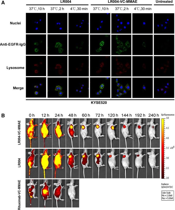
Figure 4.

Confocal analysis for intracellular localization and fluorescence imaging in KYSE520 model. (A) The internalization and lysosomal localization of LR004 and LR004‐VC‐MMAE in the KYSE520 cells by laser scanning confocal microscope. The KYSE520 cells were treated with 5 μg·mL −1 LR004 and LR004‐VC‐MMAE at 4 °C for 30 min or at 37 °C for 2 and 10 h. The lysosomes were labeled with a LAMP‐1 antibody followed by an Alexa Fluor 555‐labeled goat anti‐rabbit IgG (H+L) antibody. The cell nuclei were stained with DAPI. (B) In vivo fluorescence imaging of LR004 and LR004‐VC‐MMAE in KYSE520 nude mice xenograft model. Mice in the three DyLight 680‐labeled groups (LR004, LR004‐VC‐MMAE, and rituximab groups) were injected via the tail veins with the dose of 20 mg·kg−1 each. Representative in vivo fluorescence imaging at the indicated time points. Color scale represents photons/s/cm2/steradian.
