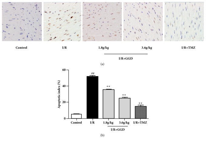
Figure 3.
Effect of GGD treatment on cardiomyocyte apoptosis in each group as detected by TUNEL assay. (a) Representative TUNEL staining of samples from rat ventricles subjected to different treatments. Apoptotic cardiomyocytes were stained brown, whereas TUNEL-negative cells were stained blue. Photomicrographs were taken at ×400 magnification. (b) Percentages of apoptotic cardiomyocytes. Values were presented as mean±SEM. n=6; #P<0.05 and ##P<0.01 compared with control group; ∗P<0.05 and ∗∗P<0.01 compared with I/R group.

