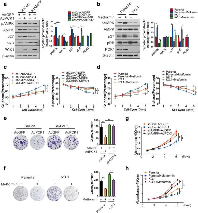
Fig. 4.
PCK1-mediated AMPK activation and delayed cell proliferation depend on p27Kip1 expression and phosphorylation of Rb protein. a, b Protein levels of the indicated proteins in PCK1-OE and KO hepatoma cells. β-actin was used as a loading control. a AMPK-stable knockdown SK-Hep1 cells (SK-Hep1/shAMPK) or control cells (SK-Hep1/shControl) were infected with AdPCK1 or AdGFP for 48 h, respectively. b PCK1-KO or parental PLC/PRF/5 cells were treated with 5 mM metformin for 12 h. Cell lysates were then subjected to immunoblot assays with the indicated antibodies for detecting the protein expression levels of p27Kip1, pRB, AMPK, and pAMPK. Results shown are representative samples from at least three independent experiments. Integrated density of these proteins was quantitatively analyzed and the results normalized to the shCon+AdGFP or parental group. c, d Flow cytometric analysis. Cells were treated as described above, collected at different timepoints (1, 2, 3, 4, or 5 days), and subjected to flow cytometry. Percentages of cells in the G1 and S phases are shown within each graph. Data represent the mean ± SD of three independent experiments. *P < 0.05. e–h Cell proliferation curves and colony formation assay. SK-Hep1 cells infected with shAMPK or shControl lentivirus were treated as described in a while PCK1-KO and parental PLC/PRF/5 cells were treated with 0.5 mM metformin. e, f Representative images and quantification of colony formation. Cell colonies were counted after 2 weeks of incubation. g, h Cell proliferation curves. Cells were counted every 24 h in triplicate. Values represent the mean ± SD of three independent experiments. *P < 0.05, **P < 0.01

