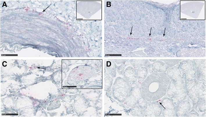
Fig. 6.
PrPCWD deposition in the kidney and salivary glands of clinically-affected white-tailed deer. a Kidney from a wt/wt deer and a (b) S96/wt deer showing periarterial PrPCWD deposition in arcuate arteries (arrows). Inserts show the specific location of these arteries in the histopathological sample. c Salivary gland from a wt/wt deer showing positive PrPCWD immunolabeling in the interstitial tissue between acini (arrows). PrPCWD immunolabeling was also detected in the ganglion neurons immersed in the salivary gland sample (insert picture). d Salivary gland from a S96/wt deer. Positive immunolabeling was detected in the same location as for wt/wt deer (arrow). No PrPCWD deposition was observed in the kidneys or salivary glands of deer expressing the H95-PrPC

