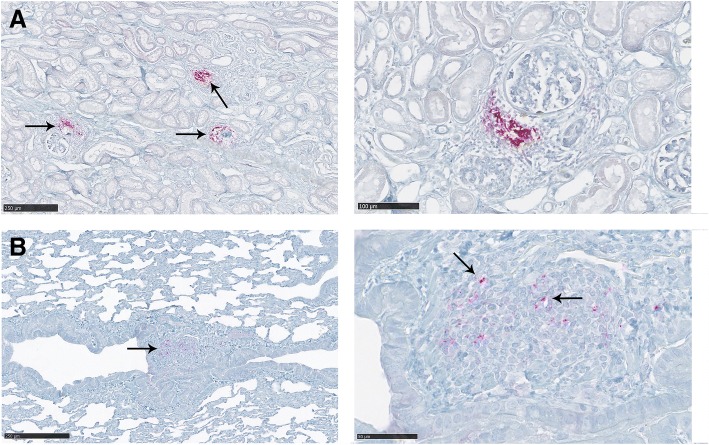
Fig. 7.
PrPCWD deposition in the kidney (a) and the lung (b) of a deer presenting signs of inflammation (D1). a A strong PrPCWD deposition associated with foci of inflammatory cells was observed in this deer. Those foci of inflammation were generally found in the proximity of the glomeruli. b A moderate interstitial inflammation was also detected in this animal. PrPCWD accumulation was observed associated with inflammatory cells (arrows)

