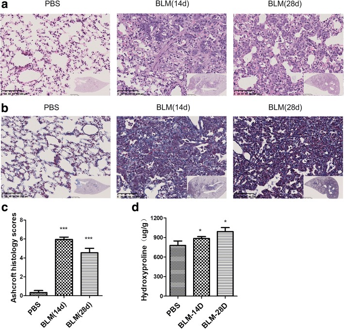
Fig. 3.
Histopathological changes of lung tissue in guinea pigs on days 14 and 28 after BLM administration. a H&E staining shows pulmonary inflammation, fibrosis, and integrity of the structure (original magnification× 20). b Masson’s staining shows deposition of collagens in lung tissues (original magnification× 20). c and d Degree of pulmonary fibrosis was evaluated by Ashcroft fibrotic scores and hydroxyproline (n = 6~7). *p < 0.05, ***p < 0.001 vs. PBS group

