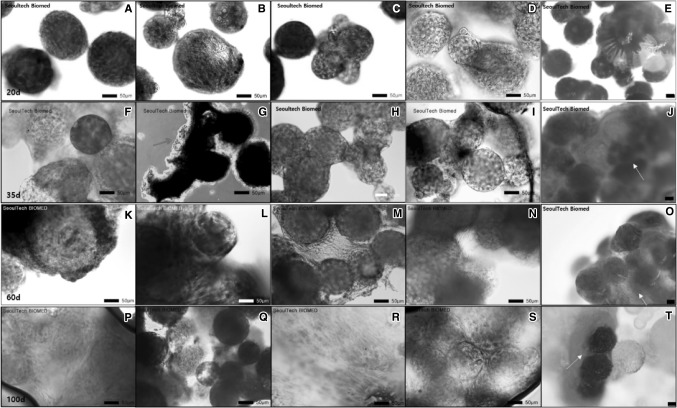
Fig. 4.
Histological stain was introduced to observe hMSC differentiation on gelatin micro-carrier according to dynamic culture condition, where hematoxylin and eosin (H&E) (A, F, K, P; × 200), MT (B, G, L, Q; × 200), Alcian blue (C, H, M, R; × 200), ALP (D, I, N, S; × 200) and Alizarin red S (E, J, O, T; × 80) stain. The yellow arrows indicate the results of Alizarin red S staining, which indicates mineralization of the regenerated tissues by Ca++ ions. The scale bar represents 50 μm. (Color figure online)

