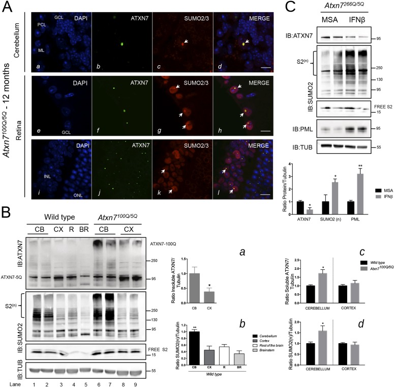
Fig. 5.
In vivo investigation of mutant ATXN7, SUMO2/3 and PML protein expression and colocalization in two SCA7 KI mouse models. (A) Cerebellum (a-d) of a 12-month-old Atxn7100Q/5Q mouse was labeled with a polyclonal anti-ATXN7 antibody and a monoclonal anti-SUMO2 antibody. SUMO2 (dot in c, see arrow) colocalized strongly (arrow in d) with an ATXN7-positive intranuclear inclusion in a Purkinje cell (92.8±4.7% of colocalization; n=200 Purkinje cells counted in lobes V and VI from n=2 Atxn7100Q/5Q mice). GCL, granular cell layer; ML, molecular layer; PCL, Purkinje cell layer. Retina (e-l) of a 12-month-old Atxn7100Q/5Q mouse: SUMO2 protein (g, k, arrows indicate dots where SUMO2 is concentrated) colocalized perfectly with mutant ATXN7 inclusions in the ganglion cell layer (h, arrows) and in a subset of cells in the INL (l, arrows). GCL, ganglion cell layer; INL, inner nuclear layer; ONL, outer nuclear layer. Representative confocal images are shown. Scale bars: 10 μm. (B) Western blot analysis of brains from 12-month-old Atxn7100Q/5Q mice and their wild-type littermates. Levels of ATXN7 and poly-SUMO2 proteins are highly increased in the cerebellum of Atxn7100Q/5Q mice compared with wild-type littermates. Quantification of soluble ATXN7/tubulin and SUMO2(n)/tubulin is shown (graphs c and d; comparison of Atxn7100Q/5Q and wild-type mice). In Atxn7100Q/5Q mice, mutant insoluble ATXN7 accumulates more in the cerebellum (1.00±0.23) than in cortex (0.38±0.13) (graph a). In wild-type littermates, poly-SUMO2 protein is more abundant in the cerebellum than in the cortex, rest of the brain and brainstem (graph b). Data are mean±s.d. from n=3 independent analyses. Statistical analysis was performed using Student's t-test: *P<0.05; **P<0.01. (C) Western blot analysis of cerebellar extracts from IFN-beta-treated Atxn7266Q/5Q mice compared with MSA-treated mice. An important increase (factor of 2.55±0.25) in the ratio of polySUMO2/tubulin was detected when comparing IFN-beta-treated with MSA-treated Atxn7266Q/5Q mice. In agreement with our previously published results (Chort et al., 2013), an increase in PML levels (factor of 3.2±0.42) and a decrease in soluble ATXN7 (by 60%) are observed in Atxn7266Q/5Q mice after IFN-beta treatment compared with MSA treatment. Data are mean±s.d. from n=3 independent analyses. Statistical analysis was performed using Student's t-test: *P<0.05; **P<0.01.

