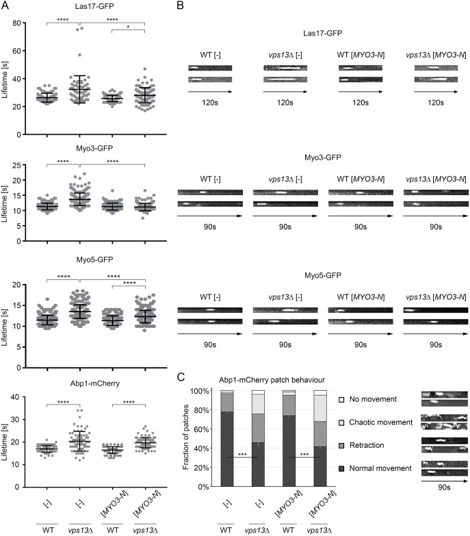
Fig. 7.
The vps13Δ mutation impairs plasma membrane events involved in endocytosis and MYO3-N suppresses some of the defects. (A) Lifetimes of Las17-GFP, Myo3-GFP, Myo5-GFP and Abp1-mCherry patches. Results were compared using one-way ANOVA followed by Tukey's multiple-comparisons test (n=87; 63; 103; 97 for Las17-GFP, n=163; 212; 207; 192 for Myo3-GFP, n=302; 264; 296; 251 for Myo5-GFP, n=83; 75; 104; 100 for Abp1-mCherry for WT [-], vps13 [-], WT [MYO3-N] and vps13 [MYO3-N], respectively); *P<0.05, ****P<0.0001. Error bars indicate s.d. (B) Kymographs of Las17-GFP, Myo3-GFP and Myo5-GFP patches. Two representative kymographs for each strain, as in A, are shown. Las17-GFP patches were observed for 120 s using an Olympus IX-81 microscope and imaging was performed via 1 s time lapse. Myo3-GFP and Myo5-GFP were observed for 90 s using an OMX DeltaVision V4 microscope and imaging was performed via 0.5 s time lapse. (C) Kymographs of Abp1-mCherry patches. Two representative kymographs for each class of patch behaviour are shown. Abp1-mCherry patches were viewed for 90 s using an Olympus IX-81 microscope and imaging was performed via 1 s time lapse. Quantification of the patches was analysed using a two-tailed Student's t-test (n=5, patches counted per replicate: 44; 45; 54; 55; 65 for WT [-], 50; 47; 60; 47; 60 for vps13Δ [-], 51; 57; 64; 37; 61 for WT [MYO3-N], 54; 69; 59; 67; 59 for vps13Δ [MYO3-N]); ***P<0.001. Statistical significances and error bars indicating s.d. are shown for patches with normal behaviour.

