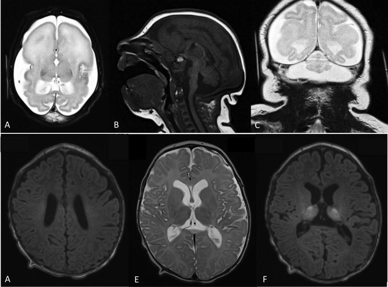
Figure 1.
(A–C) Brain MRI at age 4 days of life for patient A. A. Axial T2 shows enlarged subarachnoid spaces, anterior agyria and posterior simplified gyral pattern, enlarged lateral ventricles, absent basal ganglia and white matter hypomyelination. B. Sagital T1 shows hypoplasia of corpus callosum, brainstem and cerebellum. C. Coronal T2 shows severe hypoplasia of the cerebellar vermis and hemispheres. (D–E) Brain MRI at 3 months of life for patient B. D. Axial T1 showing hypomyelination of perirolandic white matter and corona radiata. E. Axial T2 showing age appropriate myelination of posterior limb of the internal capsule as well as heterogeneous signal in thalami and otherwise preserved brain structures. F. Axial T1 showing heterogeneous signal in thalami and otherwise preserved brain structures.

