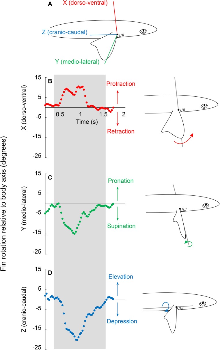
Fig. 1.
Pectoral fin rotation relative to the body axes. (A) The 3D shapes represent the body and inside fin. A joint coordinate system (FB-JCS) was placed at the proximal fin base to measure relative fin rotation about the dorso-ventral (X; red), medio-lateral (Y; green), and cranio-caudal (Z; blue) axes. (B–D) Sample rotation trace of one turn highlighted in the gray box. The pectoral fin was (B) protracted, (C) supinated and (D) depressed during the turn. This pectoral fin rotation pattern was observed in all nine trials. Protraction rotates the fin cranially, supination causes the trailing edge of the fin to translate ventrally, increasing the angle of attack, and depression makes the negative dihedral angle of the fin more negative.

