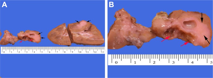
Figure 3.
Macroscopic appearances.
Notes: Gross examination of the left kidney showed that (A) the tumor was located in the mid pole of the kidney, with observable solid and cystic areas. (B) The surface of the cystic areas had tiny papillary excrescences (red arrows), and the solid area seemed grayish white (black arrows).

