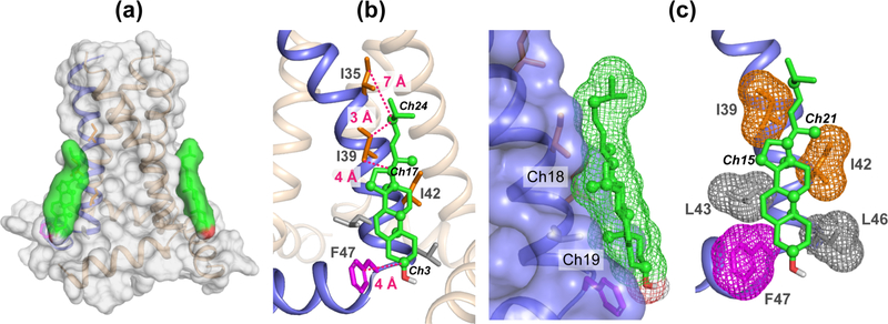
Figure 7.
Cholesterol docking onto M2 (PDB code: 2L0J) using HADDOCK. Distance restraints obtained from the 2D CC spectra were used to constrain the binding site. Cholesterol carbons that exhibit cross peaks with the M2 protein are shown as spheres. Key Ile and Phe residues at the binding interface are indicated, along with the distances to the cholesterol carbons.

