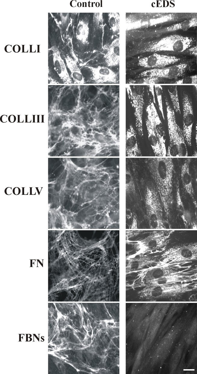
Fig 4. cEDS patients’ skin fibroblasts show the altered deposition of different structural components into the ECM.
Control and patients’ cells were analyzed with specific Abs directed against COLLI, COLLIII, COLLV, FN, and FBNs. FN and FBNs were investigated by labeling the cells with Abs recognizing all their isoforms. Images are representative of 4 control and 4 cEDS cell strains. Scale bar: 10 μm.

