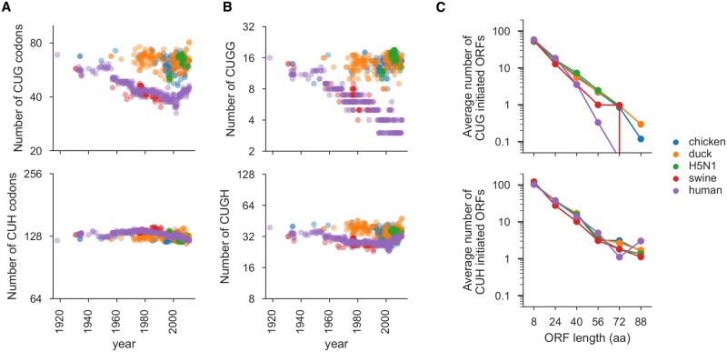
Fig 1. Evolutionary selection against CUG codons in mammalian influenza.
(A) Number of CUG (upper panel) and CUH (where H is A, U or C, lower panel) codons in reading frame 0 of viral genes from human, avian, and swine influenza sequences. X-axis indicates the year that the viral sequence was isolated. (B) Number of in-frame CUGG (upper panel) and CUGH (lower panel) motifs in reading frame 0 of the viral genes. (C) Average length in codons of CUG- and CUH-initiated ORFs in reading frames 1 and 2 of the viral genes for sequences isolated between 1970 and 2011. Lines crossing the x-axis in this panel indicate that the counts go to zero (which cannot be explicitly shown on a log scale). All plots show data only for the genes on the PB2, PA, NP, NS and M segments, since these are the segments that have not re-assorted in human lineages derived from the 1918 virus.

