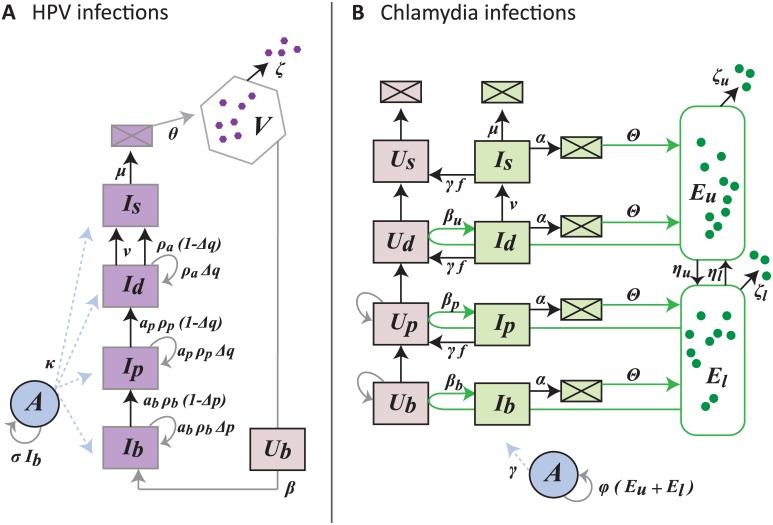
Fig 5. Flow diagram of the infection models for HPV (A) and chlamydia (B).
HPV virions, V, only infect uninfected basal cells, Ub, to become basal infected cells, Ib. Since HPV is non-lytic, infected cells follow the typical epithelial life-cycle up to the surface passing through different life stages (parbasal Ip, differentiated Id, differentiated at the surface Is). Model 3. In the case of C. trachomatis, the elementary bodies, EBs, start the infection by infecting uninfected cells in the upper layers (βu Ud Eu creates Id). The EB populations start in the upper layers, Eu, and then migrate down, ηu, into the lower layers, El. As EBs migrate down layers they enter uninfected cells (Ub and Up) and create infected cells (Ib and Ip) which die at rate α (boxes with square represent dead cells). The host immune response, A, is activated by infected basal cells in the case of HPV and all EBs in the case of C. trachomatis. Model 4. Note that for wart-associated HPV infections ρa = 0 and αb = αp = 1. See Table 3 for parameter descriptions and estimates.

