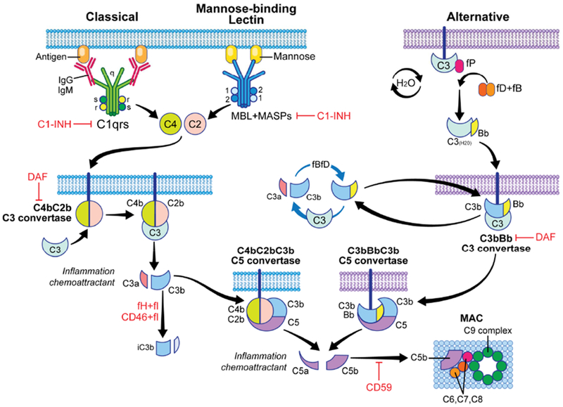
Figure 1. Overview of the complement cascade and its regulators.

Complement activation can be initiated by the classical pathway triggered by cross-linking cell-bound subclasses of IgG and IgM antibodies, the mannose binding lectin (MBL) pathway triggered by carbohydrates present on bacteria surface, and the alternative pathway that undergoes spontaneous activation on cell surfaces. All three pathways converge into one key amplification step to form multimeric C3 convertases which cleave C3 to C3a and C3b, the latter forming additional C3 convertases (amplification) and then initiating formation of the C5 convertase. Subsequently, C5 cleavage yields C5a and C5b, ultimately forming the membrane attack complex (MAC, C5b-9) on the target cells. Complement activation/amplification is restrained on self-cells by several membrane-bound and soluble regulatory proteins (shown in red). See text for further details. Surface-expressed regulators include decay accelerating factor (DAF or CD55, accelerates the decay of cell-surface assembled C3 convertases), membrane cofactor protein [MCP or CD46, co-factor for factor I (fI) that inactivates C3b to iC3b], and CD59 (protectin, inhibits formation of the MAC). Factor H (fH) is a soluble complement regulator that exhibits both decay accelerating and co-factor activity. C1 inhibitor (C1-INH) inhibits C1qrs and MBL-MASP complexes, limiting classical pathway and MBL pathway activation, respectively. MASP: mannose-associated serine protease.
