
Figure 6.
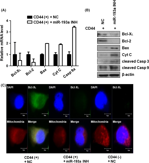
Expression of pro‐ and anti‐apoptotic genes downstream of SRSF2 in CD44(+) and microRNA (miR)‐193a‐3p‐inhibited CD44(+) cells. A, Real‐time PCR analysis of Bcl‐ XL, caspase 9a, Bax, cytochrome C and Bcl‐2 in negative control or miR‐193a‐3p inhibitor‐transfected CD44(+) MKN45 cells. Expression of the pro‐ and anti‐apoptotic genes related to SRSF2 was normalized by β‐actin and is presented as the relative ratio. B, Western blot analysis of Bcl‐XL, Bcl‐2, Bax, cytochrome C, cleaved caspase 3 and cleaved caspase 9 in negative control or miR‐193a‐3p inhibitor‐transfected CD44(+) MKN45 cells. C, Immunofluorescence assay to examine the expression of Bcl‐XL (green) in negative control or miR‐193a‐3p inhibitor‐transfected CD44(+) MKN45 cells. Mitochondria were stained with MitoSOX (red) and nuclei were counterstained with DAPI (blue). Cyt C, cytochrome C; cleaved Casp 3, cleaved caspase 3; cleaved Casp 9, cleaved caspase 9; miR‐193a INH, miR‐193a‐3p inhibitor; NC, negative control