
Figure 4.
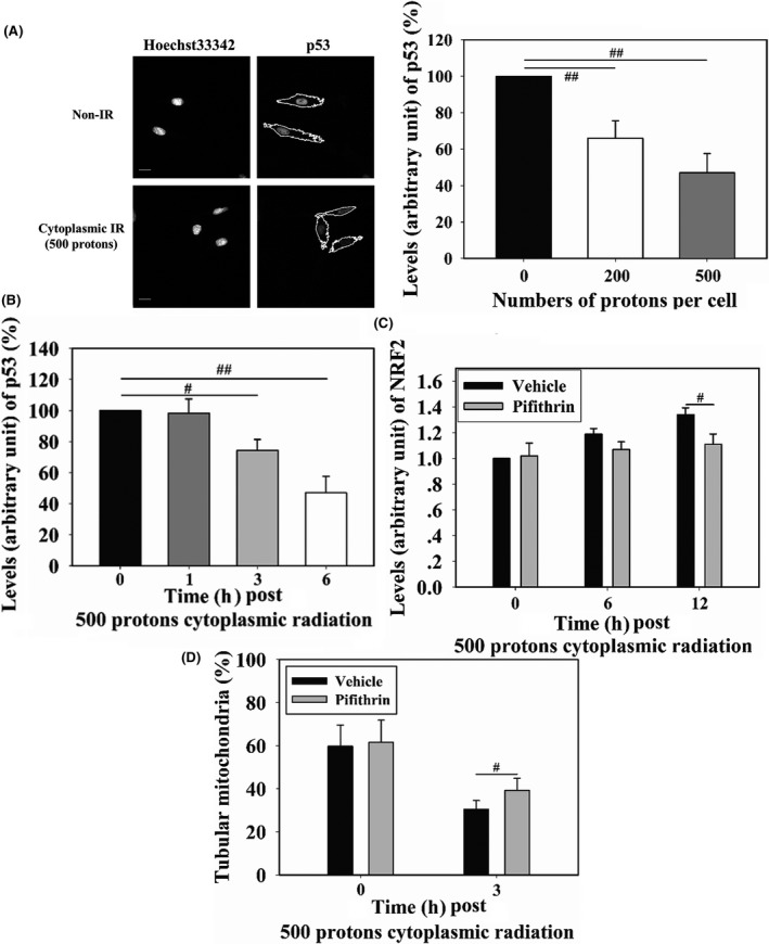
WI‐38 cells were cytoplasm targeted by 200 or 500 protons 6 h before measuring p53 levels (A). Fluorescence intensity was normalized to that of non‐irradiated cells. Cells were cytoplasm‐targeted by 500 protons and p53 levels were detected at different time points post‐irradiation (B). Effects of 20 μmol/L pifithrin‐α (p53 inhibitor) on cytoplasm irradiation‐induced nuclear factor (erythroid‐derived 2)‐like 2 (NRF2) localization to the nucleus (C) and mitochondrial morphology changes (D) were detected. Scale bar, 20 μm. IR, irradiation. # and ## indicate significant differences at P < .05 and P < .01, respectively