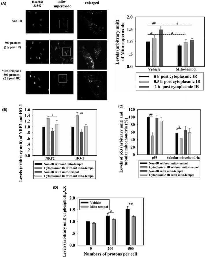
Figure 5.

WI‐38 cells were cytoplasm‐targeted by 500 protons and, at different time points post‐irradiation, mitochondrial superoxide levels were detected in cells treated with 10 μmol/L mitoTEMPOL (scavenger of mitochondrial superoxide) or non‐treated (A). Fluorescence intensity was normalized to that of non‐irradiated cells. Effects of mitoTEMPOL treatment on the levels of nuclear factor (erythroid‐derived 2)‐like 2 (NRF2) and heme oxygenase‐1 (HO‐1) (B) and on p53 expression and tubular mitochondria (C) were detected in cells targeted by 500 protons. Scale bar, 20 μm. (D) WI‐38 cells were treated with 10 μmol/L mitoTEMPOL or mock‐treated, then cytoplasm‐targeted by 200 or 500 protons. Four hours later, γH2A.X formation was detected. IR, irradiation. # and ## indicate significant differences at P < .05 and P < .01, respectively
