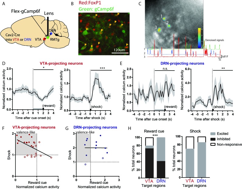
Figure 4. VTA-projecting RMTg neurons preferentially show valence-encoding patterns.
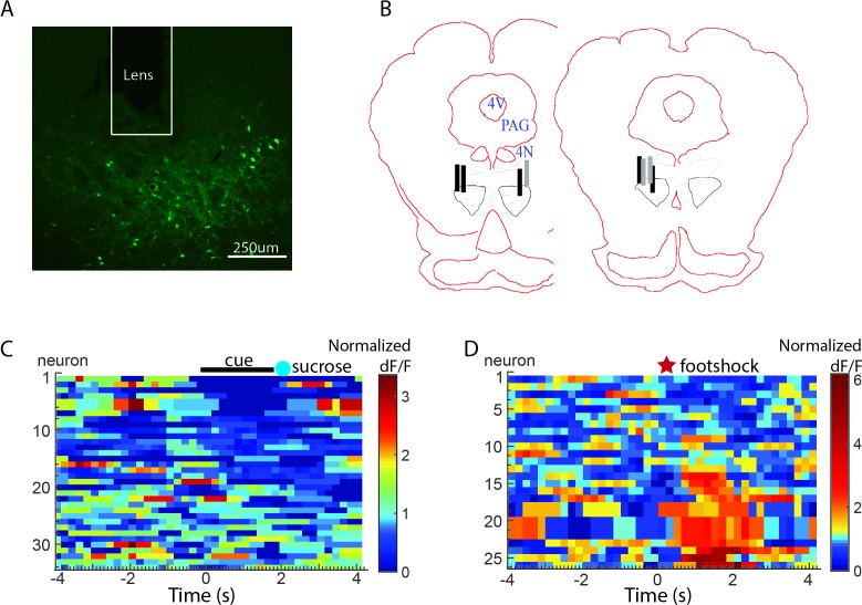
(A) Cav2-cre injected into the VTA or DRN was retrogradely transported to the RMTg in mice, driving gCaMP6f expression in subsets of RMTg neurons projecting to the VTA or DRN respectively. (B) Representative photograph of the RMTg region in which gCaMP6f in RMTg (green label) is co-expressed with FOXP1 (red), a transcription factor locally specific to RMTg neurons. (C) A representative photo of gCaMP6f positive neurons in vivo (upper panel) and denoised Ca2+ traces extracted from the marked neurons (lower panel). (D) VTA-projecting RMTg neurons showed an average inhibition by reward cues, and excitation by footshocks. (E) DRN-projecting neurons showed no average response to reward cues, but were excited by footshocks. (F) Among VTA-projecting RMTg neurons, neurons showing stronger excitations to shock tended to also show stronger inhibitions to the reward cue, while individual DRN neurons did not show this correlation (G). Colored solid dots: neurons significantly responding to both reward cues and footshocks. (H) VTA-projecting neurons were much more likely to be inhibited by the reward cue than DRN-projecting neurons, and much less likely to be activated, while VTA- and DRN-projecting neurons were both predominantly activated by shock.


