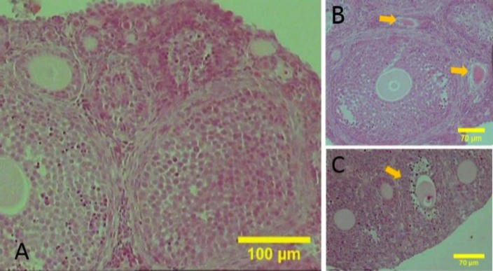
Fig. 1.
The sections of the juvenile mice ovaries stained with haematoxylin and eosin. (A) The follicles are observed in different developmental stages, (B) Follicles in late stage of atresia are located at the adjacent antral follicle (yellow arrows), and (C) Antral follicle in early stage of atresia (yellow arrows) positive, but with less intensity (Fig. 5B).

