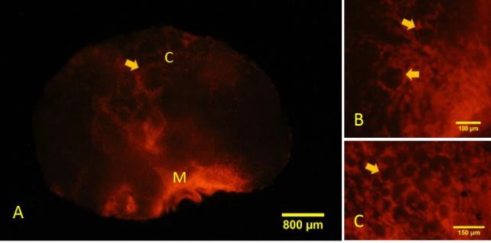
Fig. 6.
Whole-mount staining of the ovary reveals areas of propidium iodide (PI) absorption in the cortical and medullary regions. (A) Necrosis has happened mainly in the medullary region. M: Medulla, and C: Cortex. The arrow shows follicle in cortical region, (B) Somatic cells (arrow) of primary follicles are stained with PI, and (C) Somatic cells in the primordial follicles (arrow) also show necrosis

