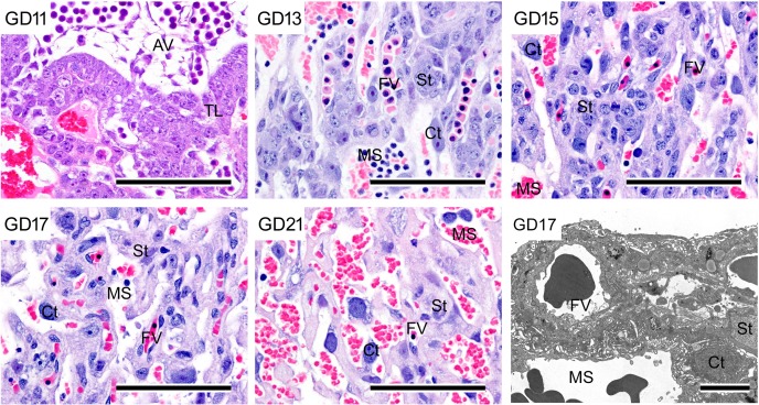
Fig. 6.
Morphological development and ultrastructure of trophoblastic septa in the labyrinth zone. Decrease in cellular density in trophoblastic septa and increase in size of cytotrophoblasts with pregnancy progression; HE stain. Bar=100 µm. Lower right, ultrastructure of trophoblastic septa. Bar=15 µm. AV, allantoic vessel; Ct, cytotrophoblast; FV, fetal vessel; MS, maternal sinusoid; St, syncytiotrophoblast; TL, trophectoderm layers.

