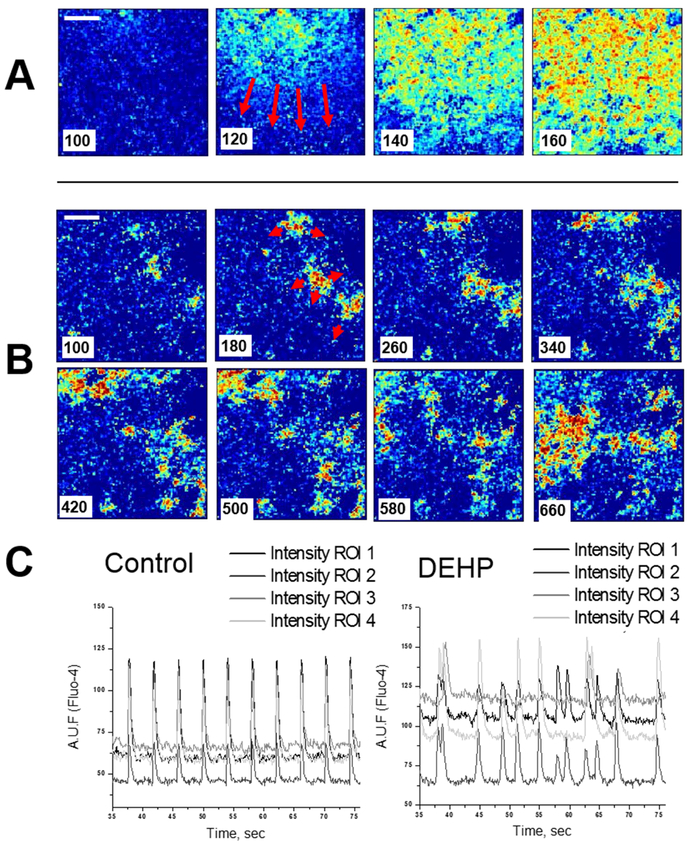
Figure 3.
Monolayers of neonatal rat cardiomyocytes display a marked decrease in conduction velocity after 3-day treatment with 50μg/ml DEHP. Individual frames from fast camera system show fast, homogenous wavefronts in control (A) versus slow, fractionated propagation in samples treated with 50μg/ml DEHP for three days (B). Note that the sequential frames in the upper and lower rows are 20ms apart, versus 80 ms for the DEHP-treated sample. A time stamp was placed on the left lower corner of the image. A ten-fold decrease in apparent conduction velocity was observed in the DEHP-treated samples. (C). Recordings of calcium transients are shown as relative units of Fluo-4 fluorescence (n=8). Reproduced with permission from26.

