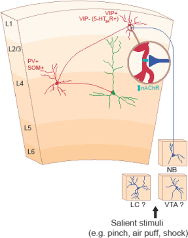
Figure 1. Circuit mechanisms of cortical disinhibition.

In layer 1 (L1) of the auditory cortex, vasoactive intestinal peptide positive (VIP+) and VIP– neurons that express the 5-HT3A receptor (5-HT3AR+) disinhibit downstream pyramidal neurons by inhibiting parvalbumin (PV+) or somatostatin (SOM+) neurons. Salient stimuli such as air puffs and shock activate neuromodulatory inputs, such as acetylcholine from the nucleus basalis (NB) that impinge upon L1 VIP+ or VIP– neurons. These inputs signal through nicotinic acetylcholine receptors (nAChR) to drive this disinhibitory circuit. Future work is required to determine whether other neuromodulatory inputs such as norepinephrine from the locus coeruleus (LC) and dopamine from the ventral tegmental area (VTA) also activate this circuit. Red denotes inhibitory neurons; green denotes excitatory neurons; and blue denotes neuromodulatory inputs. This figure is adapted from [48;53;54;57].
