
Figure 1. Levels of cardiac autoantibodies in subjects from the different groups.
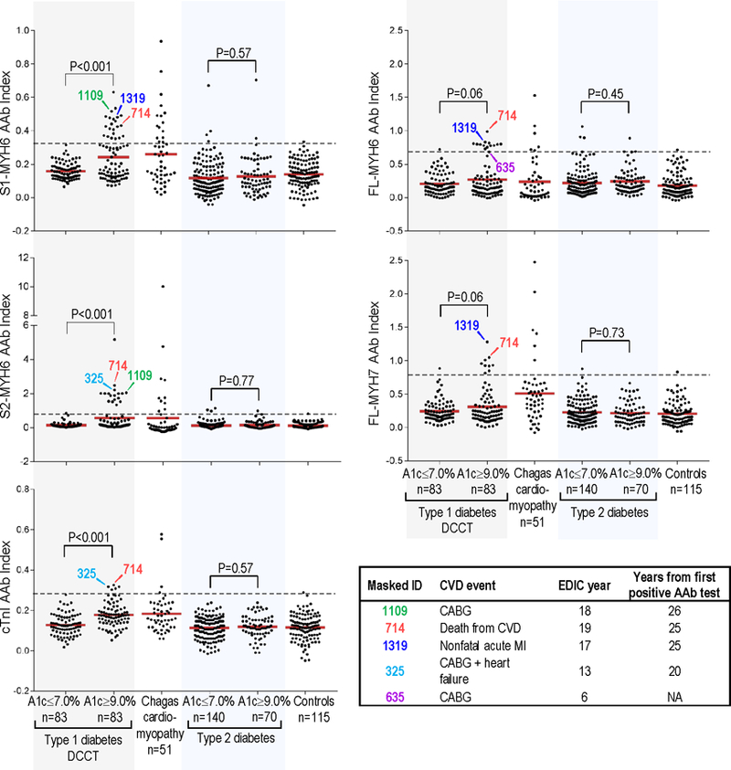
AAb, autoantibody; A1c, glycosylated hemoglobin level; dotted lines, 99th percentile cutoffs for AAb positivity. AAb levels for the T1D patients are the mean AAb index during DCCT. S1-MYH6, S1 fragment of cardiac α-myosin heavy chain (MYH6); S2-MYH6, S2 fragment of MYH6; cTnI, cardiac troponin I; FL-MYH6, full-length-MYH6; and FL-MYH7, full-length β myosin heavy chain. Controls, healthy control subjects. Inset: Subjects who subsequently had CVD events during EDIC. Number below “Masked_ID” is the DCCT/EDIC masked identifier (ID) number; CVD, cardiovascular disease; MI, nonfatal myocardial infarction; CABG, coronary artery bypass graft; NA, not applicable.