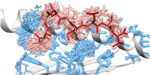
Figure 4. Side view of a pMHC complex after ensemble refinement.
Alternative conformations of selected MHC side chains are depicted in sticks, as well as alternative conformations of the ligand. All alternative conformations are compatible with the x-ray experimental data, and were obtained through a procedure of ensemble refinement (157). The single peptide conformation displayed in the crystal structure is represented in a darker shade of grey. Part of the conformational “frame” of the binding site can also be observed (i.e., a lateral alpha-helix and a floor of beta-sheets, both depicted in cartoon). Graphics were obtained with UCSF Chimera (158).

