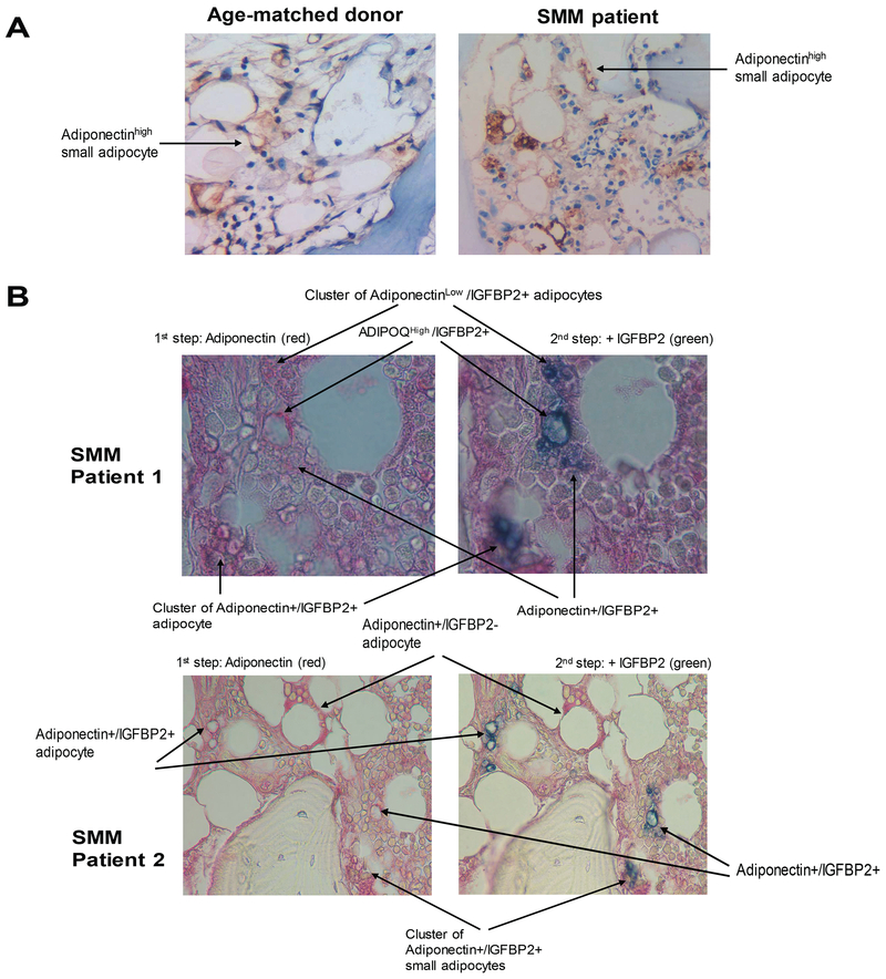
Fig 5. A subset of BM adipocytes co-express IGFBP2 and adiponectin.
(A) Immunohistochemical staining of adiponectin (stained brown) in bone sections from a healthy donor and a patient with smouldering multiple myeloma (SMM). (B) Sequential immunohistochemical staining for adiponectin (stained red) and IGFBP2 (stained green) in bone sections from patients with SMM. Note that cells positive for both markers are dark blue (red + green merging colour). Images were obtained with an Olympus BH2 microscope equipped with a 160 x/0.17 numerical aperture objective; original magnification, 200×. Images were acquired with a SPOT2 digital camera and were processed with Adobe Photoshop version 11.

