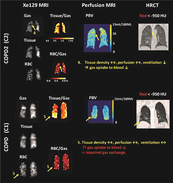
Figure 3.

Representative CT images, PBV maps, and Xe129 tissue-to-gas and RBC-to-gas ratio maps from subject C1 and C2 showed that Xe129 MRI could uniquely detect low gas exchange to blood by either low gas ventilation (area 4) or potentially impaired gas exchange from lung tissue to the blood (area 5). This information was not accessible by perfusion MRI or CT.
