Figure 1.
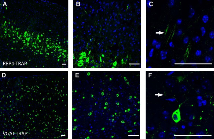
Immunofluorescence of RBP4-TRAP and VGAT-TRAP lines shows expected cellular expression patterns and localization of ribosomal protein L10a-GFP fusion to neurites. A, RBP4-driven Cre line expresses the TRAP construct, designed to tag ribosomes with GFP, in layer 5 pyramidal neurons (10×). B, Labeling extends into primary dendrites that continue into the upper layers of the cortex (40×). C, An example of a RBP4-TRAP dendrite with GFP-tagged ribosomal proteins (arrow). D, VGAT-driven Cre line expresses the TRAP construct in pattern consistent with interneurons of the cortex (10×). E, Images at a higher magnification (40×) highlight that the GFP-tagged ribosomal proteins localize in neurites. F, An example of the neurites of a single VGAT-TRAP neuron with GFP-tagged ribosomal proteins (arrow). Green, GFP; blue, DAPI nuclear stain. Scale bars, 50 μm.

