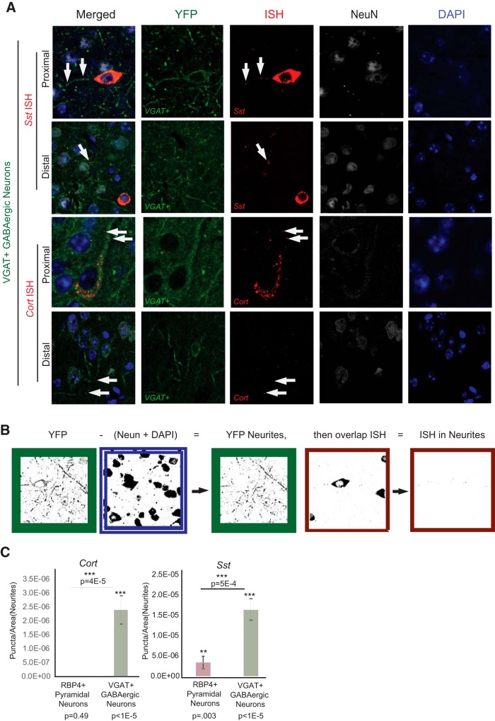
Figure 11.
In situ hybridization validation of neuropeptide mRNA in neurites. A, In situ hybridization shows RNA localization for Sst and Cort messages with immunohistochemistry of Cre-dependent membrane-bound YFP-channel fusion in each cell type. DAPI is used to label nuclei, and NeuN to define the nuclear compartment and perinuclear cytoplasm of all cortical neurons. White arrows indicate examples of Cort and Sst ISH puncta overlapping with both proximal and distal neurites in subsets of VGAT neurons. B, Illustration of the method used to quantify the ISH signal in the neurites of each cell type. The cell-specific Cre-driven YFP signal (green border) was masked with NeuN and DAPI (blue and white border) to remove any signal in nuclear and perinuclear compartments. The remaining ISH puncta overlapping with GAP (red borders) were quantified. C, Quantification of overlapping ISH puncta with cell-specific labeling of neurites reveals that Cort and Sst are significantly enriched in neurites of VGAT neurons. Each probe and no probe control, n = 13, Mann–Whitney test. **P < 0.01, ***P < 0.001.

