
Figure 1.
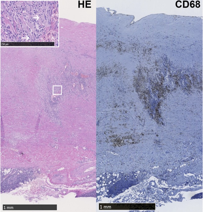
LV-GCA aorta with granulomatous inflammation and giant cells. Representative image of Hematoxylin Eosin (HE) and CD68+ macrophages in the media layer of the aorta from a LV-GCA patient. The white box shows magnified giant cells (white arrows) in the HE staining.