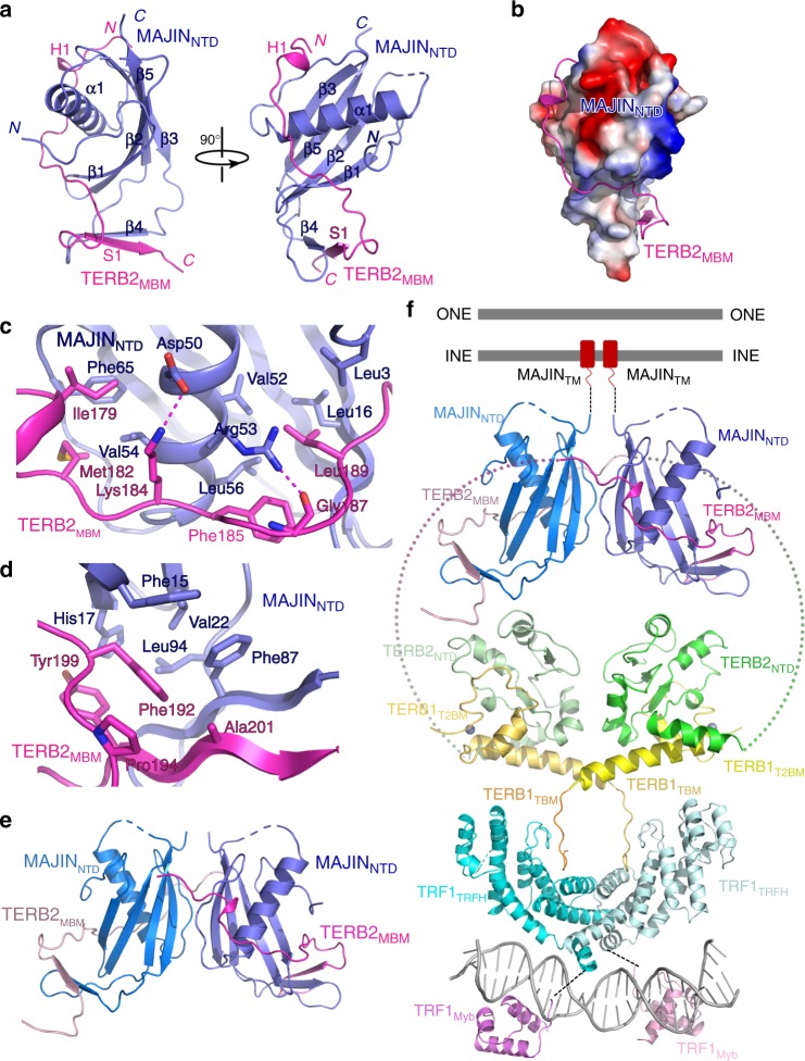
Fig. 3.
Crystal structure of the TERB2MBM–MAJINNTD complex. a Ribbon diagrams of two orthogonal views of the TERB2MBM–MAJINNTD complex. TERB2MBM is colored in magenta and MAJINNTD in blue. b Electrostatic surface potential of TERB2MBM-binding site of MAJINNTD. Positive potential, blue; negative potential, red. c, d Detailed interactions at the TERB2MBM–MAJINNTD interface. The color scheme is the same as in a. Residues important for the interaction are shown in stick models. Salt bridges and hydrogen-bonding interactions are shown as magenta dashed lines. e Ribbon diagram shows the potential heterotetramer mode of the TERB2MBM–MAJINNTD complex revealed in the crystal structure. f Structural model of the connection between telomeres and the NE through the TRF1–TERB1–TERB2–MAJIN interaction network

