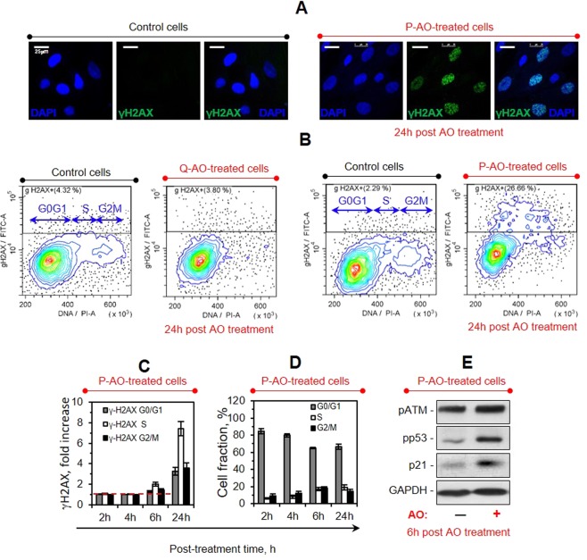
Figure 4.
Treatments with antioxidants induce replicative stress in P-AO-treated cells. (A) Immunofluorescence analysis of the P-AO-treated eMSCs reveals γH2AX foci accumulation within 24 h post AO treatment, scale bar = 25 μm. (B–D) Flow cytometry studies of the γH2AX + cell distribution among the cell cycle phases in the control, P-AO-treated and Q-AO-treated cells performed at different time points after the cell treatments. Representative dotplots of cell cycle distributions of the eMSCs stained with γH2AX antibody (B), mean γH2AX immunofluorescence signals in G0/G1, S and G2/M cell fractions normalized to the control values (C) and corresponding cell cycle distributions (D) are shown. The data show slow accumulation of γH2AX foci in cells replicating their DNA, which is correlated with the decelerated S phase progression in P-AO-treated eMSCs. Mean values ± SD of three independent experiments are shown. (E) Western blot analysis of the DDR-related protein (pATM, pp53, and p21) expression in P-AO-treated eMSCs after 6-hour AO treatment. The blot was stained with Ponceau S Red and then cut at the appropriate molecular weights of proteins of interest. Abbreviations: eMSCs, endometrial mesenchymal stem cells; γH2AX phosphorylated histone H2AX; pATM, phosphorylated ATM kinase; pp53, phosphorylated p53; p21, cyclin-dependent kinase inhibitor p21waf1/cip1; AO, antioxidant Tempol at 2 mM concentration; P-AO-treated cells, eMSCs exposed to 2 mM of Tempol at 14 h post-serum stimulation; DDR, DNA damage response; SD, standard deviation.

