Figure 1.
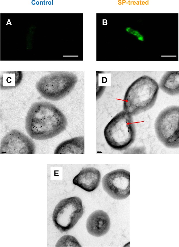
Effect of SP on the expression of immunoreactive EfTu at the surface of B. cereus. EfTu was localized by CLSM (A,B, Scale Bar = 2 µM) and electron microscopy (C–E Scale Bar = 100 nm) in control (left part) and SP treated bacteria (right part) using EfTu polyclonal antibodies. For CLSM, immunolabeling was realized in the absence of permeabilization procedure. For electron microscopy, EfTu was localized on ultrathin section by the immunogold technique. Arrows indicate EfTu immunoreactive material localized at the bacterial periphery. E: Control view realized by exposing bacteria to EfTu polyclonal antibodies preincubated with P. aeruginosa EfTu (10−6 M).

