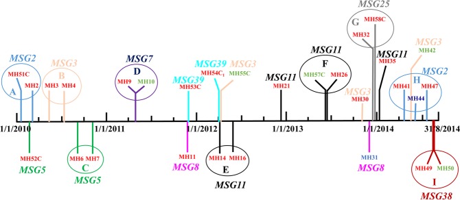
Figure 3.
Color-coded timeline showing the distribution of 29 C. parapsilosis isolates exhibiting cluster fingerprinting patterns obtained from 29 patients in NICUs of the Maternity Hospital in Kuwait. The patient number (MH1, MH2, MH3 etc.) yielding C. parapsilosis at the indicated time points (vertical colored lines) are shown along with the corresponding cluster microsatellite genotype (MSG) in bold and italicized letters of same color. The location of patients in the four NICUs is indicated by patient number color: red font, patients in NICU-1; green font, patients in NICU-3 and blue font; patients in NICU-4. Colonizing strains are indicated by letter “C” after patient number. The patients yielding isolates belonging to same MSG that were recovered within 62 days of each other are shown in circles (color coded with each MSG) marked ‘A’ to ‘I’.

