Figure 2.
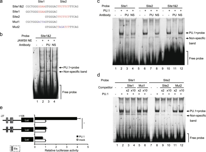
Identification of the PU.1 binding site. (a) Probes used in EMSAs. (b) The FLO-labeled Site 1&2 was incubated with the nuclear extracts prepared from JAWSII cells in the presence of either anti-PU.1 (PU) or non-specific (NS) Abs. (c) The FLO-labeled indicated probes were incubated with recombinant PU.1 protein in the presence of either anti-PU.1 (PU) or non-specific (NS) Abs. (d) The FLO-labeled indicated probes were incubated with recombinant PU.1 protein in the presence of 2-fold (×2) or 10-fold (×10) amounts of non-labeled identical WT or mutated competitor. After electrophoresis in 5% acrylamide gels, fluorescence was detected. (e) 293 T cells were transfected with either empty (mock) or PU.1-expression (PU.1) plasmids together with either WT or mutant reporter plasmids described in Fig. 1. At 48 h after transfection, luciferase and β-galactosidase activities were measured. Luciferase activities were normalized to β-galactosidase activities. Data are expressed as the ratio of the luciferase activity of the respective promoter-less plasmid-transfected cells. Results are shown as means + S.D.s (n = 3). Similar results were obtained in three independent experiments. *p < 0.05. Full-length gels with lower contrasts for (b–d), are included in Supplemental Information.

