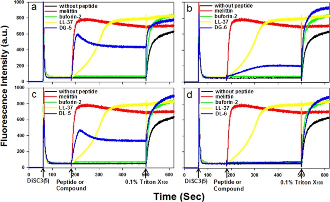
Figure 5.
Depolarization effects of compounds (DG-5 (a); DG-6 (b); DL-5 (c) and DL-6 (d)) and peptides on S. aureus cytoplasmic membrane. The membrane potential-sensitive fluorescent dye 3,3′-dipropylthiadicarbocyanine Iodide (DiSC3(5)) was used for determining the S. aureus membrane depolarization by measuring fluorescence of the dye release (excitation λ = 622 nm, emission λ = 670 nm). Except for melittin (2 μM), the concentration of control antimicrobial peptides and antimicrobial compounds used is 2X MIC against S. aureus (KCTC 1621) (buforin-2 (16 μM); LL-37 (5 μM); DG-5 (5 μM); DG-6 (5 μM); DL-5 (5 μM), DL-6 (5 μM)). LL-37 and melittin were used as a positive control peptides and buforin-2 was used negative control peptide. The other two triplicate images are presented in Fig. S4 (SI).

