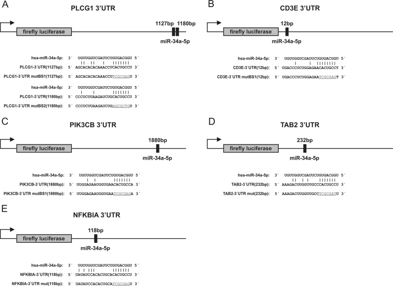
Fig. 1. Schematic representation of reporter gene constructs.
The position of the predicted miR-34a-5p binding sites in the respective 3′UTR reporter constructs and additionally the sequences of the binding sites of miR-34a-5p in the different 3′UTRs as well as the mutated binding sites (underlined) are shown. a PLCG1-3′UTR reporter vector, b CD3E-3’UTR reporter vector, c PIK3CB-3′UTR reporter vector, d TAB2-3′UTR reporter vector, f NFΚBIA-3′UTR reporter vector

