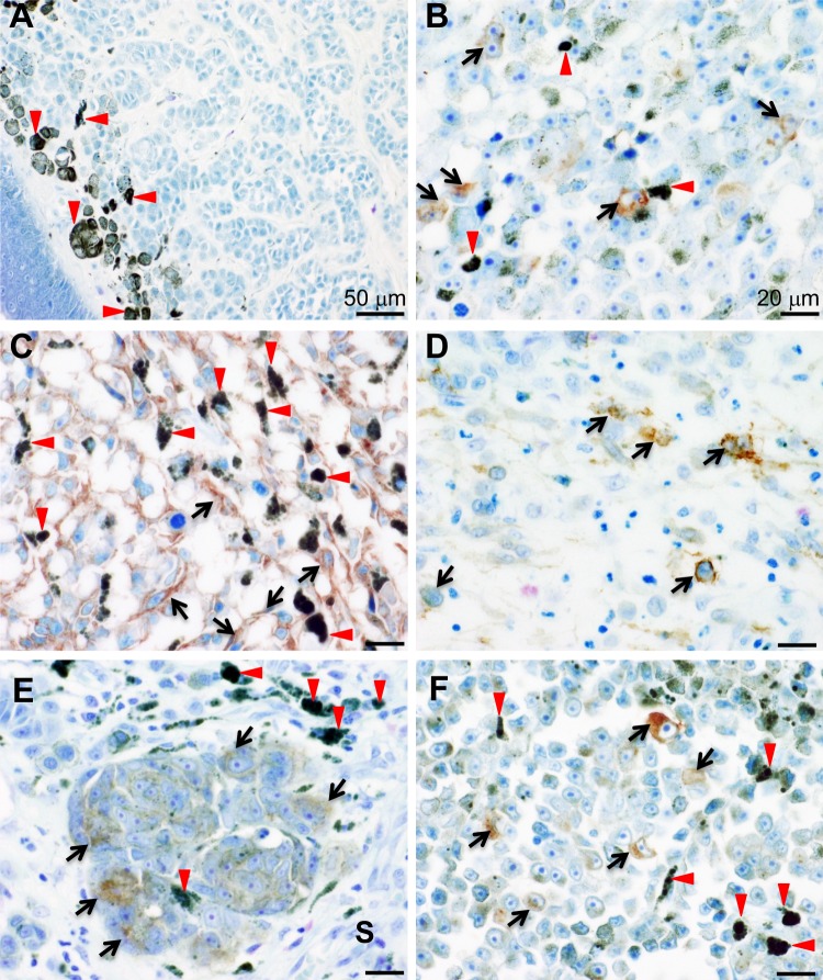
Figure 1.
Tissue microarray analyses for IL13Rα2 expression. Multiple series of tissue microarrays were subjected to immunohistochemical analysis by using anti-IL13Rα2 antibody (KH7B9). Expression of IL13Rα2 was detected in the cytoplasm or membrane of melanoma cells (arrows). Red arrowheads indicate melanin pigment. (A) Benign naevus of the right face. (B) Metastatic malignant melanoma from the armpit (lymph node). (C) Malignant melanoma of the thigh. (D) Malignant melanoma of the cunnus. (E) Malignant melanoma of the skin. IL13Rα2 was expressed by melanoma cells (arrows) but not by stromal cells (S). (F) Malignant melanoma of the right sole. Scale bar: 50 μm (A), 20 μm (B–F).

