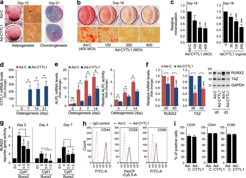
Fig. 3. Overexpression of CYTL1 inhibits the osteogenesis of hMSCs.
a Representative staining images of the adipogenic and chondrogenic differentiations of hMSCs (n = 5). b Representative staining images of the osteogenic differentiation of hMSCs infected with Ad-C or Ad-CYTL1 (n = 10). c Quantitation of Alizarin red S staining in hMSCs infected with Ad-C or Ad-CYTL1 (left, n = 10) or treated with recombinant CYTL1 protein (right, n = 8). d, e The CYTL1 mRNA level (d, n = 7), ALPL enzyme activity, and ALPL mRNA level (e, n = 5) in hMSCs (day 0) and osteogenically differentiating hMSCs infected with Ad-C or Ad-CYTL1. f mRNA and protein levels of RUNX2 and TAZ in hMSCs (day 0) or osteogenically differentiating hMSCs (day 3) infected with 400 MOI of Ad-C or Ad-CYTL1 (n = 6). g RUNX2 reporter gene activity in hMSCs (day 0) or osteogenically differentiating hMSCs on days 3 or 7 after transfection with empty vector (EV, 1 μg) or expression vectors encoding mouse Runx2 (1 μg) or mouse Cytl1 (amounts indicated, μg) (n = 5). h, i Expression of hMSC markers (CD44, CD29, and CD90) in hMSCs infected with 400 MOI of Ad-C or Ad-CYTL1 (n = 4). Data represent the means ± SEM of the indicated number of independent experiments; *p < 0.05, **p < 0.005, and ***p < 0.0005, as determined by one-way ANOVA (c, g) or two-tailed t-test (d, e, f, i)

