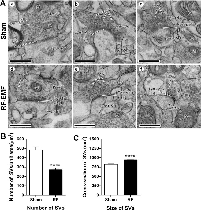
Figure 3.
Changes in the number and size of SVs in mouse striatum after RF-EMF exposure. (A) Representative TEM micrographs of the synaptic region in the striatum were acquired from sham- (a, b and c) and RF-EMF-exposed (RF) mice (d, e and f), respectively. M, mitochondria; Pre-SN, pre synaptic neuron; Post-SN, post synaptic neuron; SVs, synaptic vesicles; scale bars, 500 nm. (B,C) Comparison of SV density and size between sham- and RF-EMF exposed mice. 30–31 synapses were randomly chosen in each condition. (B) Shown are numbers of SVs per square micron (C) Shown are the cross-sectional areas (nm2) of the SVs. Each bar represents the mean ± SEM. Statistical significance was evaluated using two-tailed unpaired Student’s t-test (****P < 0.0001).

